Cách đặt avatar mặc định Facebook, tránh lộ ảnh cá nhân
Cách đặt ảnh avatar Facebook về mặc định có thể thực hiện trên máy tính, điện thoại nhanh chóng. Tổng hợp những hình avatar mặc định độc đáo trên Facebook. Xem ngay nhé!
Cách để avatar mặc định Facebook giúp bạn hạn chế công khai hình ảnh của bản thân trên mạng xã hội. Cách chỉnh ảnh avatar Facebook về mặc định có thể thực hiện vô cùng nhanh chóng trên các thiết bị. Xem ngay bài viết để không bỏ lỡ những thông tin hữu ích nhé!

Lợi ích khi đặt avatar mặc định Facebook
Ảnh đại diện mặc định trên Facebook sẽ xuất hiện sau khi bạn tạo tài khoản Facebook thành công. Tuy nhiên, nhiều người vẫn giữ nguyên hay đặt avatar Facebook về mặc định, vì các lý do như:
- Tránh công khai hình ảnh cá nhân trên mạng xã hội khi không cần thiết.
- Thuận tiện hơn trong việc sử dụng tài khoản phụ cho các hoạt động như seeding, công việc,...
- Ảnh đại diện mặc định Facebook có thể khiến tài khoản trở nên khác biệt hơn.

Do vậy, cách để ảnh đại diện mặc định trên Facebook càng được nhiều người quan tâm và tìm kiếm. Phần tiếp theo sẽ hướng dẫn chi tiết về cách thực hiện trên các thiết bị vô cùng đơn giản.
Cách để avatar mặc định trên Facebook trên điện thoại, máy tính
Ảnh đại diện mặc định trên Facebook có thể được thao tác nhanh chóng trên điện thoại và máy tính. Dưới đây là hướng dẫn chi tiết cho cách thực hiện trên 2 thiết bị này.
Cách chỉnh avatar Facebook về mặc định trên điện thoại
Cách đặt ảnh đại diện mặc định trên Facebook được thực hiện trên điện thoại như sau:
Bước 1: Bạn mở và đăng nhập vào ứng dụng Facebook của mình trên điện thoại. Sau đó, bạn nhấn vào biểu tượng Cá nhân như hình dưới đây. Trong giao diện Trang cá nhân, hãy chọn vào nút biểu tượng Avatar cá nhân.

Bước 2: Tiếp theo, trong hộp thoại hiện ra, bạn chọn Xem ảnh đại diện. Khi hình ảnh hiện ra, bạn nhấn vào biểu tượng ba chấm ở phía trên, góc phải màn hình.

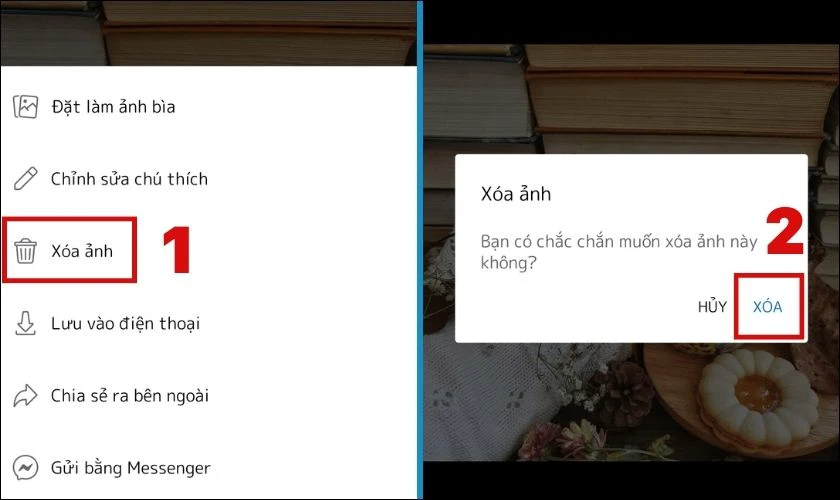
Bước 3: Trong hộp thoại này, bạn nhấn vào Xóa ảnh và chọn Xóa để xác nhận thao tác. Cuối cùng, bạn có thể khởi động lại ứng dụng lần nữa để cập nhật chỉnh sửa.

Khi đó, bạn đã thực hiện xong cách chỉnh ảnh avatar Facebook về mặc định. Sau đó, bạn có thể thay đổi ảnh avatar mới bất kỳ lúc nào bạn muốn.
Cách chỉnh avatar Facebook về mặc định trên máy tính
Bên cạnh điện thoại, bạn cũng có thể chỉnh ảnh đại diện Facebook về mặc định trên máy tính. Cách để avatar mặc định trên Facebook bằng máy tính như sau:
Bước 1: Bạn truy cập vào trang web Facebook trên máy tính theo đường link https://www.facebook.com/. Trong giao diện Facebook, bạn nhấn vào biểu tượng Cá nhân như hình bên dưới.

Bước 2: Lúc này, bạn nhấn vào biểu tượng Avatar cá nhân. Trong hộp thoại xuất hiện, bạn chọn Xem ảnh đại diện.

Bước 3: Trong giao diện mới, bạn nhấn vào biểu tượng ba chấm và chọn Xóa ảnh.

Bước 4: Sau đó, bạn chọn nút Xóa để hoàn tất thao tác.

Như vậy, bạn đã thực hiện xong cách chỉnh ảnh avatar Facebook về mặc định bằng máy tính.
Sưu tầm ảnh avatar mặc định trên Facebook đổi nền độc đáo
Sau khi đặt avatar mặc định Facebook thành công, bạn có thể lựa chọn đổi một avatar mới cho chúng. Bên cạnh ảnh đại diện, bạn có thể đặt một avatar hoạt hình để hiển thị trên trang cá nhân của mình. Đặc biệt, avatar hoạt hình này sẽ được tạo thành dựa trên khuôn mặt của bạn.
Lưu ý: Cách tạo avatar nhân vật hoạt hình chỉ có thể được thực hiện bằng điện thoại.
Các bước tạo avatar hoạt hình trên Facebook như sau:
Bước 1: Bạn mở ứng dụng Facebook trên điện thoại và nhấn vào biểu tượng Trang cá nhân. Sau đó, bạn chọn nút Chỉnh sửa trang cá nhân.

Bước 2: Kế đó, bạn nhấn vào nút Tạo avatar. Trong giao diện chụp hình, bạn căn chỉnh khuôn mặt vào khung tròn theo hướng dẫn trên màn hình. Sau đó, bạn nhấn vào biểu tượng Máy ảnh ở phía dưới màn hình để chụp lại.

Bước 3: Tiếp theo, bạn lựa chọn màu da cho nhân vật hoạt hình của mình và chờ avatar này được tạo thành.

Bước 4: Cuối cùng, trên màn hình sẽ hiện ra nhân vật hoạt hình của bạn. Nếu không còn chỉnh sửa gì thêm, bạn có thể nhấn vào Xong để hoàn tất thao tác.

Lúc này, trong trang cá nhân của mình, bạn có thể vuốt qua ở biểu tượng Ảnh đại diện để thấy được avatar vừa tạo thành. Nếu muốn chỉnh sửa, hãy nhấn vào biểu tượng Cây bút ở avatar hoạt hình và thực hiện các điều chỉnh theo ý muốn.
Ngoài ra, bạn cũng có thể sử dụng một số ảnh đại diện được thiết kế lại từ mạng để đặt avatar cho mình. Dưới đây là các kiểu avatar trên Facebook giúp trang cá nhân của bạn trở nên độc đáo hơn.
Ảnh đại diện mặc định trên Facebook ảnh chế


Ảnh đại diện mặc định trên Facebook nền nhiều màu


- Link tải: https://drive.google.com/drive/folders/1ID69xd005wqqYuixtVkgfYr9cBEscMe7?usp=sharing
Đây là một số những sưu tầm về những avatar Facebook độc đáo. Với những hình ảnh trên, chắc hẳn bạn cũng đã lựa được một hình avatar thật ngầu rồi đúng không. Hãy lưu lại ngay nhé!
Giải đáp thắc mắc khi đặt ảnh đại diện mặc định trên Facebook
Nhiều người vẫn còn một số thắc mắc xoay quanh cách chỉnh ảnh avatar Facebook về mặc định. Câu trả lời sẽ được trình bày ở phần dưới đây.
Có cách để ảnh đại diện mặc định Facebook là nữ hay nam không?
Trước đây, ảnh đại diện mặc định trên Facebook sẽ được phân biệt cho nam và nữ. Chúng sẽ được đặt dựa trên thông tin cá nhân của bạn trên Facebook.
Tuy nhiên, hiện nay, ảnh đại diện mặc định Facebook sẽ được dùng chung cho cả nam và nữ. Cách để avatar mặc định trên Facebook đã được trình bày ở những phần trên. Bạn có thể tham khảo thử nhé!
Có thể tương tác với ảnh avatar mặc định Facebook không?
Khi để avatar mặc định Facebook, người khác không thể tương tác với bài viết về việc chỉnh sửa này. Thông thường, sau khi đặt avatar, bài viết về ảnh đại diện mới sẽ xuất hiện trên newsfeed của bạn. Và người khác có thể nhìn thấy và tương tác với chúng.

Tuy nhiên, với ảnh đại diện mặc định, Facebook sẽ không hiện bài viết trên trang cá nhân của bạn. Do vậy, người khác chỉ thấy ảnh đại diện mặc định mà bạn chỉnh sửa nhưng không thể tương tác.
Nếu bạn muốn có được avatar mặc định nhưng vẫn hiện bài viết, bạn có thể download avatar trên mạng giống avatar mặc định. Sau đó, bạn đặt chúng thành ảnh đại diện của mình. Như vậy, bài viết sẽ được hiện trên trang cá nhân của bạn và người khác có thể tương tác với chúng.
Kết luận
Trên đây là hướng dẫn chi tiết về cách đặt ảnh avatar mặc định Facebook đơn giản. Hi vọng những thông tin trên có thể giúp bạn chỉnh avatar của mình về mặc định nhanh chóng nhất. Đừng quên chia sẻ bài viết nếu thấy chúng hữu ích bạn nhé!
Bạn đang đọc bài viết Cách đặt avatar mặc định Facebook, tránh lộ ảnh cá nhân tại chuyên mục Thủ thuật ứng dụng trên website Điện Thoại Vui.
Admin
Link nội dung: https://pi-web.eu/cach-dat-avatar-mac-dinh-facebook-tranh-lo-anh-ca-nhan-1735868123-a2845.html