Tổng Hợp 1001+ Ảnh Bia Rượu Tự Chụp Đẹp Cực Chill
Hãy cùng Spicy Food Studio tổng hợp ảnh rượu bia tự chụp cực đẹp bao chill và lan tỏa niềm văn hoá nhậu văn minh qua bài viết dưới đây nhé.
Từ rất lâu rồi rượu bia luôn là thứ không thể thiếu trong những cuộc vui nhậu nhẹt tới bến! Hãy cùng Spicy Food Studio (chuyên chụp ảnh food, chụp hình quảng cáo sản phẩm, chụp hình cafe, chụp ảnh sữa chua đẹp, chụp menu quán ăn, chụp hình bánh ngọt…) tổng hợp ảnh bia rượu tự chụp cực đẹp bao chill và lan tỏa niềm văn hoá nhậu văn minh qua bài viết dưới đây nhé.
Rượu bia có lợi ích gì?
Nếu chỉ uống rượu bia với một lượng vừa phải, thì thức uống này rất tốt. Nhiều chuyên gia bác sĩ đã khuyên rằng nên được sử dụng rượu bia mỗi ngày với một lượng theo chỉ định đối với những người có sức khoẻ ổn định.

Lợi ích của bia rượu
1 chai bia trung bình sẽ có lưu lượng khoảng 330ml tương đương với 110ml rượu vang và 30ml các loại rượu nặng. Đây là lượng bia rượu vừa đủ cho một ngày để đảm bảo an toàn và sức khỏe cho người dùng. Sử dụng rượu bia vừa đủ sẽ mang lại lợi ích sau:
- Kích thích vị giác giúp ăn ngon, tiêu hoá tốt, dễ dàng hấp thụ dưỡng chất cho cơ thể
- Tăng cường lưu thông máu dễ dàng hơn, hiệu quả.
- Giảm nguy cơ mắc bệnh về tim mạch nhờ tăng HDL cholesterol tốt trong mạch máu.
- Giảm stress, giúp tinh thần thoải mái và dễ đi vào giấc ngủ hơn.

Ảnh chụp cocktail đẹp
Rượu bia là thức uống ngon không thể thiếu trên mỗi bàn tiệc lớn nhỏ, cho những buổi gặp mặt thân mật thêm thân tình. Và chắc chắn rằng những dịp gặp gỡ này không thể bỏ qua hình ảnh bia rượu tự chụp phải không nào.
Ảnh bia rượu tự chụp đẹp nhất
Những bức ảnh bia rượu tự chụp sẽ luôn là những hình ảnh tự nhiên nhất và lưu giữ nhiều kỷ niệm quý giá nhất đối với mỗi người. Sau đây là cách chụp ảnh với chai bia ly rượu mà các bạn có thể tham khảo.

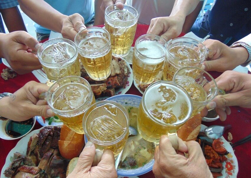
Hình chụp cụng ly rượu với nhau

Rượu tạo ra niềm vui cho các buổi tiệc

Một cốc bia tươi cho tinh thần sảng khoái

Ảnh bia rượu tự chụp tại nhà

Hình ảnh uống bia buồn ở quán nhậu

Hình ảnh bia rượu đẹp

Không khí buổi tiệc thêm náo nhiệt cùng các ly bia

Nâng chén rượu cùng anh em bạn bè

Ảnh tự chụp ly rượu
Chụp ảnh chai bia rượu tại Spicy Food Studio
Ảnh bia rượu tự chụp có vẻ không quá phức tạp. Tuy nhiên nếu muốn hình ảnh rượu bia lên một tâm cao mới hãy để các nhiếp ảnh gia dày dạn kinh nghiệm thực hiện.
Vì vậy tại sao bạn không thử tìm đến Spicy Food Studio qua số hotline: 0907 289 726 (Zalo) nếu có nhu cầu chụp hình ảnh rượu bia đẹp chuyên nghiệp.

Spicy Food Studio chuyên chụp hình rượu bia cho quảng bá thương hiệu

Kết hợp chụp hình chai bia cùng phông nền xanh mát

Chụp ảnh rượu vang cho những buổi tiệc sang trọng

Hình ảnh chụp rượu mạnh

Một ly cocktail dịu nhẹ cực chill

Hình ảnh rượu vang đẹp bắt mắt

Ngày hè làm sao có thể bỏ qua ly cocktail mát lạnh

Cốc beer tươi kết hợp cùng vài lát chanh vàng và đá viên

Tonic giúp pha chế rượu thêm ngon hơn

Ảnh chụp rượu vang sang trọng
Chúng tôi là những nhiếp ảnh gia giàu kinh nghiệm đã thực hiện hàng trăm dự án chụp ảnh đồ ăn, chụp hình đồ uống… Cùng đội ngũ nhân sự nhiệt huyết, tận tâm chắc chắn sẽ mang lại những hình ảnh rượu bia đẹp giá trị nhất.
Không nên uống rượu bia khi tham gia giao thông
Rượu bia nếu được sử dụng vừa phải sẽ tốt cho sức khoẻ, nhưng khi đã uống rượu bia dù ít hay nhiều cũng không nên tham gia giao thông.
Mức phạt thổi nồng độ cồn đối với các cá nhân vi phạm luật an toàn giao thông: phạt tiền từ 2.000.000 – 8.000.000 VND. Tịch thu giấy phép lái xe từ 10 – 24 tháng.

Bữa ăn thêm trọn vẹn nhờ có những chai bia
Các hình phạt trên đều là những hình phạt rất nặng khi sử dụng rượu bia mà tham giao thông. Bởi vì uống rượu bia khi tham gia giao thông có thể để lại nhiều hậu quả nặng nề gây nguy hiểm đến tính mạng cho chính mình và những người xung quanh.
Hãy tuân thủ nghiêm túc quy định uống rượu bia thì không lái xe để tạo ra văn hoá nhậu văn minh.
Hy vọng qua bài viết , độc giả của Spicy Food Studio đã có thêm những ý tưởng mới là về hình ảnh thông qua ảnh bia rượu tự chụp cực chill mà chúng tôi cung cấp.
Admin
Link nội dung: https://pi-web.eu/tong-hop-1001-anh-bia-ruou-tu-chup-dep-cuc-chill-1735833312-a2728.html